![]()
![]()
![]()
![]()
|
|
|
|
|
The outcome of the discrimination analysis is available on the Results page.
Result Images
The large image display shows a fusion of the clustered t-maps with the normalized patient images.
Note: The cluster images are a synthetic compilation of information with values in the range from 0 to 230. The value of 230 corresponds to abnormal voxels and is shown in red using the default color table.
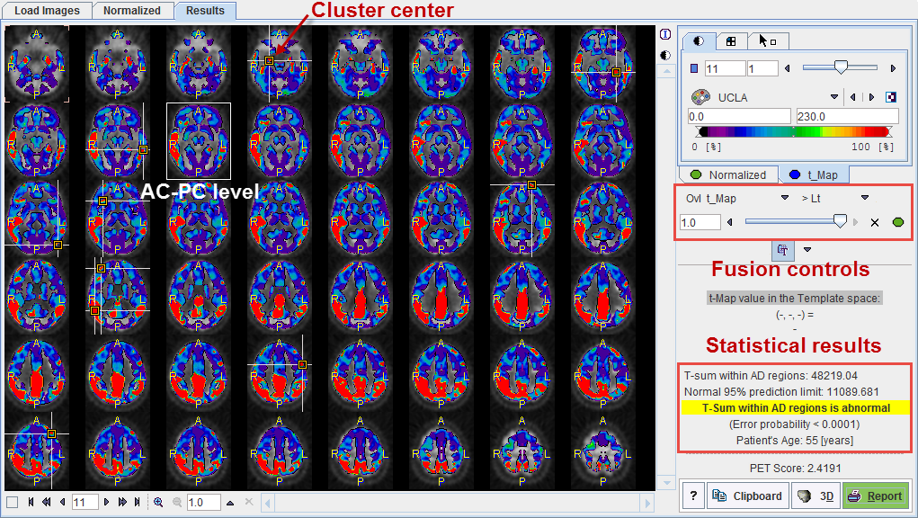
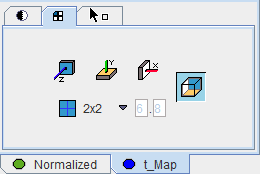
Initially, 2 mm thick slices parallel to the AC-PC line are shown in a 6x8 layout as illustrated above, but the arrangement can be changed as usual using the layout tab.
Using the fusion slider the mixture between the normalized patient images (fully to the left) and the cluster t-maps (fully to the right). There are many other fusion options available to closely examine the data, as described in the PMOD Image Fusion Guide.
The image at the AC-PC level is enclosed in a white rectangle. The cluster maxima are indicated in the images by markers and crossing lines. With the Go to button in the markers panel of the t_Map it is possible to directly jump to these markers, and also to the AC-PC plane.
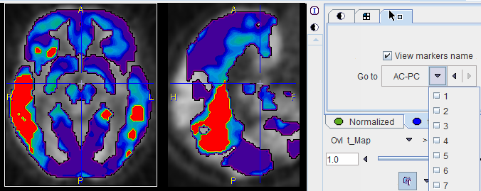
By enabling the View markers name box, the cluster numbers can additionally be shown in the image overlay.
The Template space section allows localizing the points of interest in the MNI template space. As soon as the cursor is moved over the images, the voxel coordinate is reported together with the t-value.
Note: The MNI template coordinate system is a right-handed patient-oriented system (like the Talairach system). Therefore, the x-coordinate is increasing from the right side of the image (patient left) towards the left side, which might be somewhat unexpected in the beginning.
Statistical Results
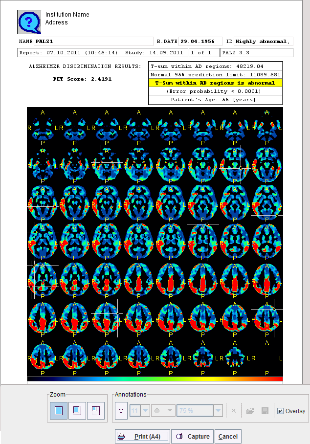
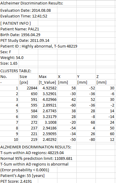
The numeric results are summarized in the lower right corner. The AD t-sum calculated for the patient (example below: 48219) is shown together with the upper normal limit (always 11089). If it is above 11089 the finding is abnormal, and the error probability is stated (p<0.0001).
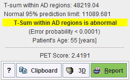
The PET score has a value of 1 for an AD t-sum equal to the normal limit, and >1 for abnormal t-sums. It has been shown to be closely related to the stage of AD, providing a measure of cognitive impairment. As it has a high test-retest reliability, it can be used as an imaging biomarker for clinical trials aiming at preventing dementia in MCI patients.
The results can be saved via the Clipboard button, and then pasting into a suitable program such as MS Excel, as illustrated below.
Results Documentation
When the Report button is activated a report dialog window appears. It contains the numeric outcome as well as the cluster images from inferior to superior, including markers at the cluster centers. The images are helpful for documenting left/right asymmetries.