The purpose of a VOI analysis is to calculate the distribution of pixel values within delineated tissue structures. There are several VOI definitions available in PMOD.
Contour VOIs
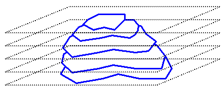
In PMOD a contour VOI is defined as a stack of planar, closed polygons which are named ROIs (region-of-interest). The contours are manually or semi-automatically outlined on the loaded images, and the pixels contained within the contour boundaries are considered for the VOI statistics.

There are a few principles to bear in mind when working with contour VOIs.
This hierarchy of three levels
is reflected in the VOI user interface. Each level has a corresponding tool bar for object manipulations. For instance, when selecting the VOI button and shifting, the contours in all slices will be shift. When selecting the ROI button and shifting, only the contours in the current slice will be shifted.
Template VOIs
A template VOI is an image file which contains numeric label information for the different anatomic structures as the image information. This VOI information can be loaded into PMOD and used for statistics. The template VOI approach has the advantage that the structures can be arbitrarily complex, and that the results of external segmentation programs can easily be used within PMOD. The disadvantage is that the VOI definition cannot be modified. However, in PMOD it is simple to convert template VOIs into contour VOIs.
Often, template VOIs are used in combination with atlas images. Brain atlas images are representative images of the (human, rat, etc.) brain imaged with a certain modality (PET, SPECT, T1 MR, T2 MR, etc.) showing a normal brain. Usually, the images of many normal subjects are brought into alignment and are then averaged. This results in somewhat blurry template images which show the characteristic pattern of the brain in the particular modality. The template images are used as a basis for the standard analysis of individual images. First, the images are spatially normalized (elastic warping) to the template, and then a set of standard VOIs is applied to the normalized images to obtain regional statistics. Typically, the VOIs in the template space are template VOIs which are universally applicable, not contour VOIs which are program-specific.
The PMOD software includes a human brain template VOI (AAL), two rat brain template VOI (Sprague-Dawley by W. Schiffer and Wistar by Tohoku), a mouse brain template VOI (M. Mirrione) and a primate brain template VOI (Cynomologus Monkey) with corresponding normalization templates in the image fusion tool. For using the template VOIs a user should first spatially normalize the individual brain images using the corresponding normalization template.
Map VOIs
A map is a file, which contains label numbers in all pixels which identify different objects in the image. PMOD can load and analyze such a file and create a VOI template out of it. A special case are the object maps (*.obj) resulting from segmentations in AnalyzeAVW. These can directly be loaded as Map VOIs.
Mask VOIs
A mask is a binary file, which contains 1 in all pixels belonging to the tissue of interest, and 0 in all other pixels. It is most likely the result of some threshold or segmentation operation of the images to be analyzed. A mask-based VOI analysis provides two pixel distributions, from the tissue of interest and from the background. Any image can be loaded and used as a mask VOI. If it is not a binary image, the user can specify the threshold below which the pixels are converted to 0, while the other pixels are converted to 1.
Starting VOI Analysis
PMOD supports the access to VOI analysis in almost all situations when an image is displayed. The VOI button in the tools list next to the image
immediately starts the VOI tool with the current images. In the PVIEW tool, there is a separate tab VOIs for entering the VOI definition mode.