![]()
![]()
![]()
![]()
|
|
|
|
|
The Crop Heart dialog window serves for creating an approximation of the LV myocardium, which will be used for initializing the contouring.
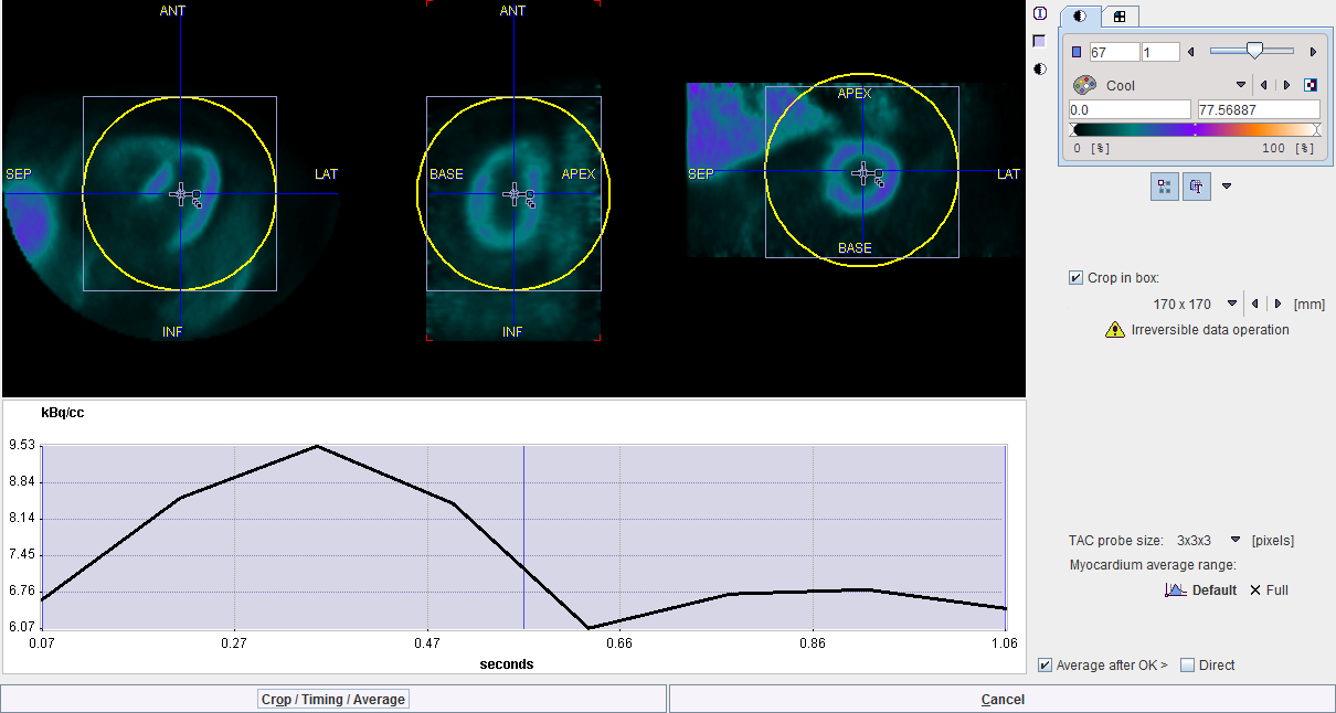
The shaded area represents the time range for the gate averaging. It can be adjusted by dragging the vertical edges. Default resets the range, and Full includes all gates. The resulting average image is shown in the image window at the top.
Enable the Crop in box in order to define a cropping volume. A yellow sphere is shown in the overlay. Its size can be changed with the list selection, and its center defined by dragging the handles. It should be placed such that the whole heart is enclosed, with enough margin for the short axis reorientation. The cropped image can be masked outside the sphere by checking the Mask box.
With each click into the image an average time-activity curve (TAC) at that location is calculated and shown in the curve panel. The sampling is performed by averaging the pixels with the configurable TAC probe size.
The action button to continue is located in the lower left. It changes the naming according to the enabled options. With all options enabled (recommended) the button is labeled Crop/Timing/Average.
Arrangement of the Result Images
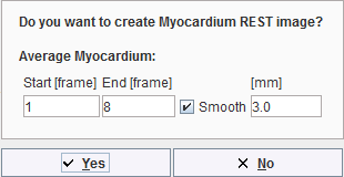
Whenever the time-averaging is performed, a new image study called MYOCARDIUM is calculated, and the display is switched to MYOCARDIUM. The user can switch between the original DYNAMIC study and the averaging results by the study selection as illustrated below. Note the red BLOOD entry which has no function in the gated analysis.
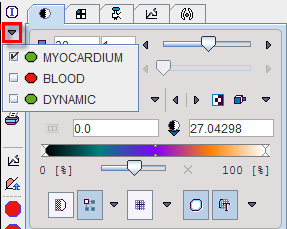
To transform the image into standard SA orientation activate the dedicated button  . If the "automatic" check for the SA reorientation is on, this step is immediately performed after averaging.