![]()
![]()
![]()
![]()
|
|
|
|
|
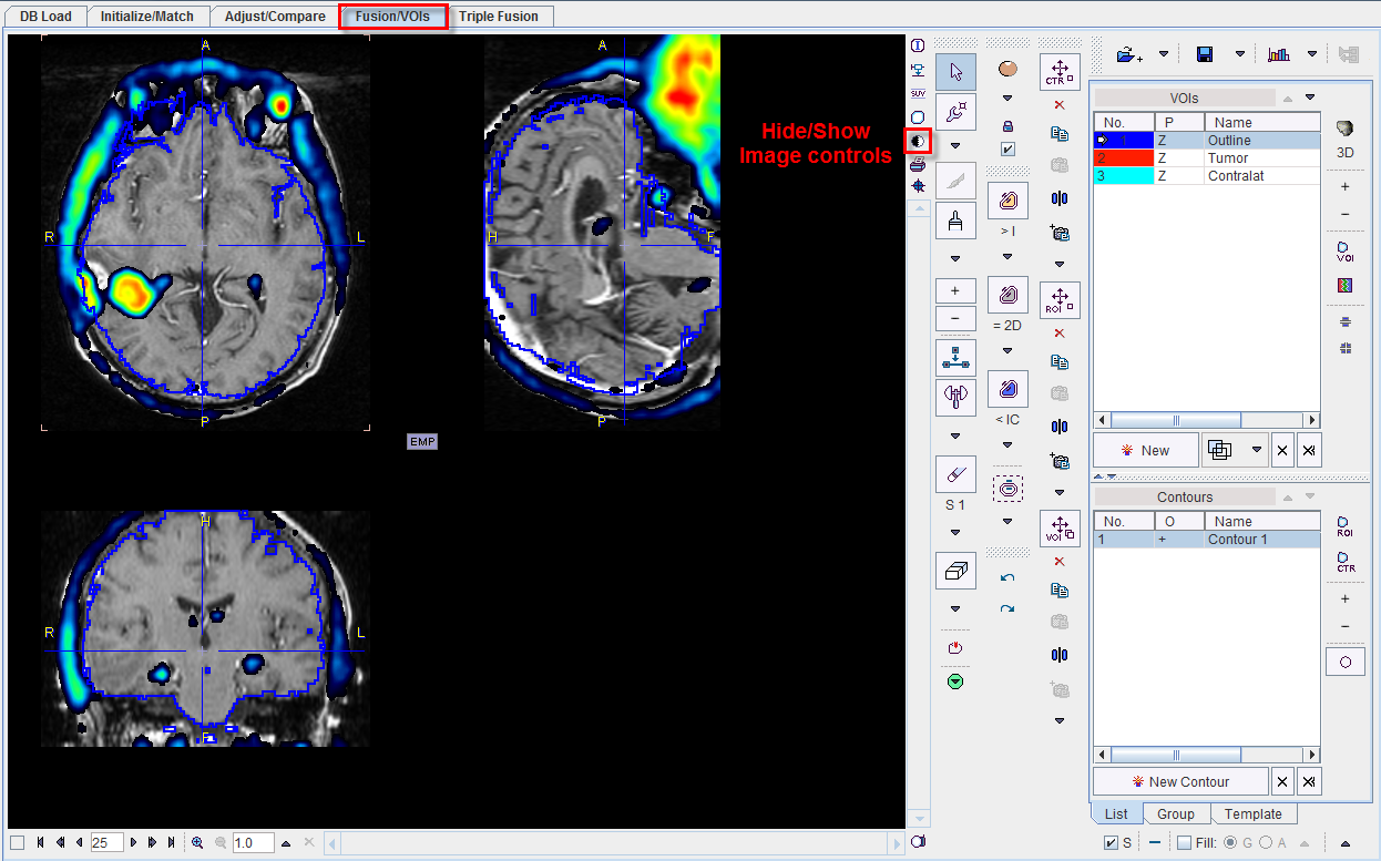
The indicated VOI button besides the image toggles between the VOI definition mode and the reviewing mode. In principle, VOI definition works exactly as described in the PMOD Base Functionality Guide. The only thing one has to care about is, that the selected Source study (A or B) is relevant for VOI definition and evaluation. So before using one of the iso-contouring or region-growing tools please select the relevant source.
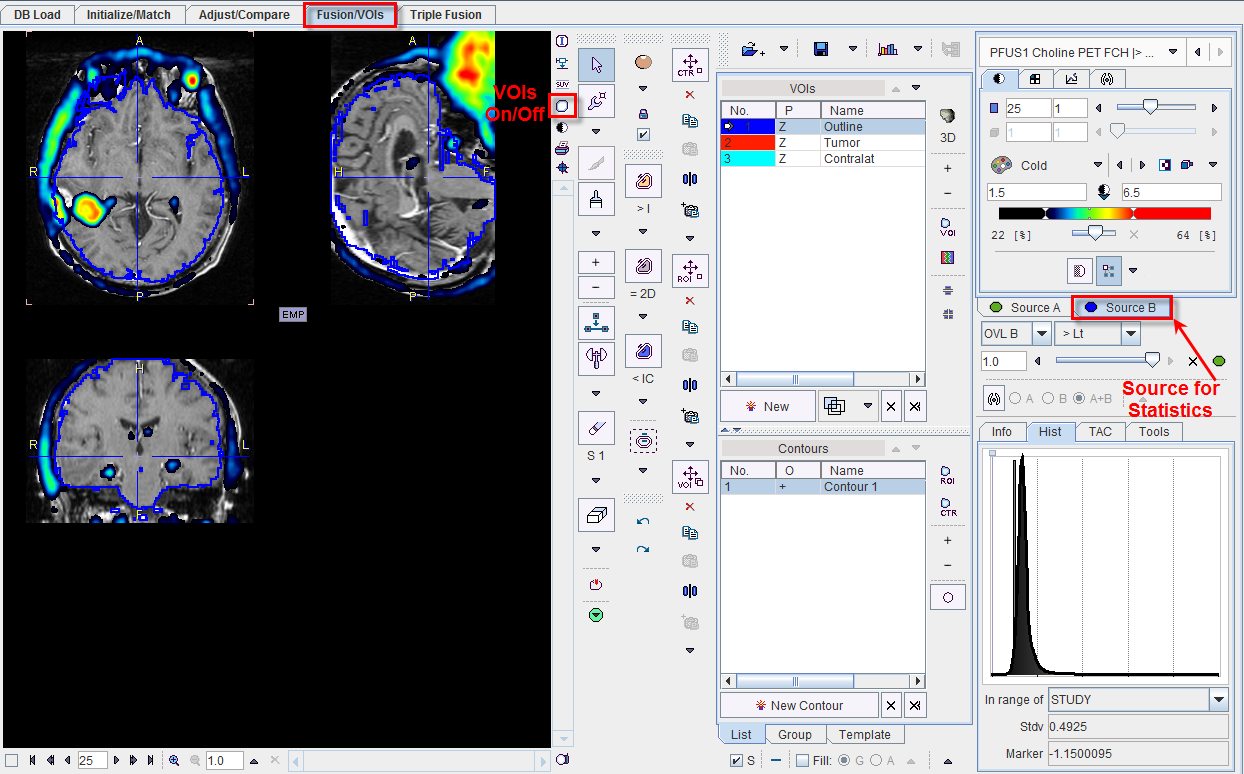
The example below shows the fusion of a MRI and a Choline PET in the VOI mode, with Choline (Source B) selected as the active study. The tumor VOI is obtained by defining a bounding box VOI and using the iso-contour auto-voi. When the Statistics button is selected, it starts the statistics calculations with the Choline study. To evaluate the statistics of the MRI, first select Source A, and then Statistics.
After a suitable configuration of the image presentation and the selection of the appropriate source the image controls can be hidden with the  button to get more image space as illustrated below. They can be brought back using again.