![]()
![]()
![]()
![]()
|
|
|
|
|
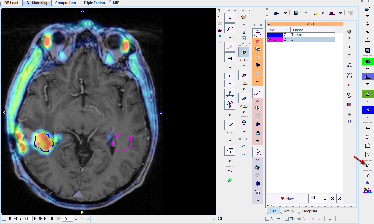
The VOIs page is illustrated below. It serves for outlining volumes-of-interest directly in the fused images.

VOI Definition and Evaluation
The standard VOI options are available for the VOI creation. Please refer to the PMOD Base Functionality Guide for explanations of the VOI functionality. The only distinctive thing to consider is, that the series selected on the tab to the right (A or B) is relevant for VOI definition and evaluation. In the example above, the choline PET series A is selected, so that the hot iso-contouring ![]() was successful in detecting the tumor boundary. When the statistics is calculated with the
was successful in detecting the tumor boundary. When the statistics is calculated with the  button, the choline uptake in the tumor uptake is obtained. Otherwise, had the tab B been selected, iso-contouring would have operated on the MRI and failed in the tumor outlining task.
Image Selection
If more than one input series has been processed or image algebra results were generated, there are several candidate images for the VOI statistics. The two selections in the lower right allow freely defining which series is configured on the A and B tabs. After a suitable configuration of the image presentation and the selection of the appropriate source the image controls can be hidden with the  button to get more image space as illustrated below. They can be brought back using again.
Action Buttons
Assuming that all input images have been registered to the reference, the user can proceed to the various post-processing pages with the two action buttons.
|
Switches to the Comparison main page for visualizing multiple fused images. |
|
Switches to the MIP main page for creating rotating fusion MIP renderings. |
Alternatively the main pages Comparison and or MIP can directly be selected with the tabs.