![]()
![]()
![]()
![]()
|
|
|
|
|
ORGAN SEPARATION by Local Means Analysis (LMA)
PSEG implements the local means analysis (LMA) method licensed from CEA, Orsay, France. This method aims at segmenting "functional organs" characterized by a particular pharmacokinetics. A functional organ only corresponds to an entire anatomical organ if the organ is functionally homogeneous. Otherwise, the functionally of different organ parts are separated and can be treated individually. The patented LMA method has been shown to be robust regarding the low signal-to-noise ratio, the limited spatial resolution and potential organ movement of dynamic rodent PET studies [1,2,3].
The LMA segmentation includes the following processing steps:
Tissue structures which cannot be isolated by the LMA segmentation because they do not kinetically differ from the neighborhood need to be outlined using the general VOI features of PMOD.
k-Means Clustering
In addition to the LMA method which incorporates prior knowledge, PSEG offers a k-means clustering as a general method for subdividing a volume into clusters of "similar" pixels [5]. The time-weighted Euclidean distance is used as the measure of dissimilarity (or distance) between TACs. In PSEG, the procedure performs the following steps for the pixels within a mask, given a prescribed number N of clusters
Note that because no geometric information is used for the clustering, spatially disconnected pixels will most likely be included in the resulting clusters.
Supervised Clustering by a Set of Kinetic Class TACs
k-means clustering is working completely data-driven without any prior knowledge apart from the number of clusters. PSEG offers a complementary supervised clustering approach where the user specifies a set of TACs which are characteristic for the kinetics of different tissues. This method was introduced by Turkheimer et al. [6] for determination of gray matter reference tissue without specific binding in [11C]PK11195 brain studies. It requires preparation of time-activity curves which are representative for different tissues in the brain. They found that the method worked best after normalizing the data frame-wise by subtracting the frame average and dividing by the frame standard deviation.
The implementation in PSEG is close to the supervised clustering variant presented by Ikoma et al. [7] for use with [11C]PIB. Particularly, the weight ratio (eq. 3 in [7]) is used to assign each pixel to one of the classes, or to background.
Note that the data has to be normalized outside of PSEG using the z-score external tool.
Partial-Volume Effect
PET images are inherently affected by the partial-volume effect. This means that the measured tracer activity concentration is not accurate due to the relatively low image resolution and the limited tissue sampling. The low spatial resolution of the PET system causes a blurring of the image, so that high activities (from a hot lesion) are spread to the surrounding as illustrated below. This effect is called spill-out. The same effect also causes a spill-in of background activity into the volume of interest.
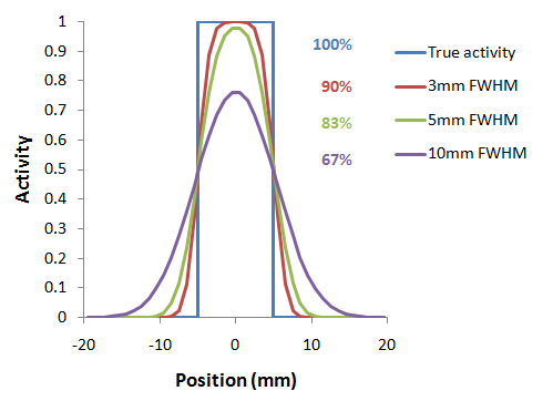
As a consequence, hot lesions tend to appear less aggressive (reduced maximum) but bigger (spreading) than they are in reality.
Partial-volume effects are complex: Spill-in and spill-out depend on the geometry of the objects, the activity distribution of the tracer, and on the resolution of the scanner which may vary across the imaging field-of-view. Therefore, practical correction approaches have to assume certain conditions and can only be approximate.
VOI Based Partial-Volume Correction (GTM Method)
The Geometric Transfer Matrix (GTM) method according to Rousset et al. [4] restricts partial volume correction to the signal of the true objects which are constituted by VOIs. The relation of measured PET values (affected by the partial-volume effect) to the true PET values is given by the matrix equation below
![]()
with the following notations:
Ctrue |
Vector of the true average activity concentration in the different VOIs of interest. The vector length n equals the number of object VOIs. |
Cmeasured |
Actually measured average activity concentration in the different VOIs. Each VOI is assumed to have a homogeneous concentration. |
GTM |
Geometric Transfer Matrix which describes the spill-over among all the VOIs. The matrix is square with nxn weighting elements wi,j which express the fraction of true activity spilled over from VOIi into VOIj. In practice, wi,j is calculated as follows: A binary map is created with 1 in all pixels of VOIi and 0 elsewhere. The map is convolved with the imaging Point-Spread Function (PSF), and in the resulting spillover map the weighted average of all VOIj pixels calculated. |
The GTM equation above represents a system of linear equations. Once the weights have been calculated, the system can be solved for the true values Ctrue by matrix inversion. It has been shown [4] that this algorithm is robust to noise propagation during the correction process.
LMA Variant of GTM Method
The LMA (Local Means Analysis) GTM method [1] uses the homogeneous regions localized by the segmentation and calculates the average uptake in the inner of the structures. The percentage of pixels per segment considered for averaging is a parameter of the method. With 100% pixels included, the LMA GTM method equals the standard GTM method.