![]()
![]()
![]()
![]()
|
|
|
|
|
The Factor Analysis (H2O, Lung TAC) model performs a factor analysis (FA) on dynamic cardiac H215O PET data to obtain anatomical images of the heart. Please refer to the guide of the cardiac modeling tool PCARD for details.
Principle
One TAC must be specified which is assumed to represent H215O activity in the lung. The model then proceeds as follows:
During model preprocessing three TACs are mathematically derived from the Lung TAC
During pixel-wise processing myocardium and blood pool images are calculated by a factor analyis:
Acquisition and Data Requirements
Image Data |
Dynamic cardiac H215O PET study (decay corrected). |
TAC 1 |
Time-activity curve representing blood activity in the lungs. |
Model Preprocessing
The lung TAC must be specified as a VOI or a file. It is then used together with the input parameters to calculate the expected TACs in the left ventricle (LV), the right ventricle (RV), and the myocardium.
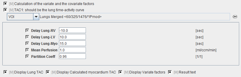
Delay Lung-RV |
Left shift of the lung TAC to the time when the bolus arrived in the RV. |
Delay Lung-LV |
Right shift of the lung TAC to the time when the bolus arrived in the LV. |
Delay Lung-Myo |
Right shift of the lung TAC to the time when the bolus arrived in the myocardium. |
Mean Perfusion |
Expected mean perfusion of the myocardium. |
Partition Coefficient |
Partition coefficient of water in myocardium |
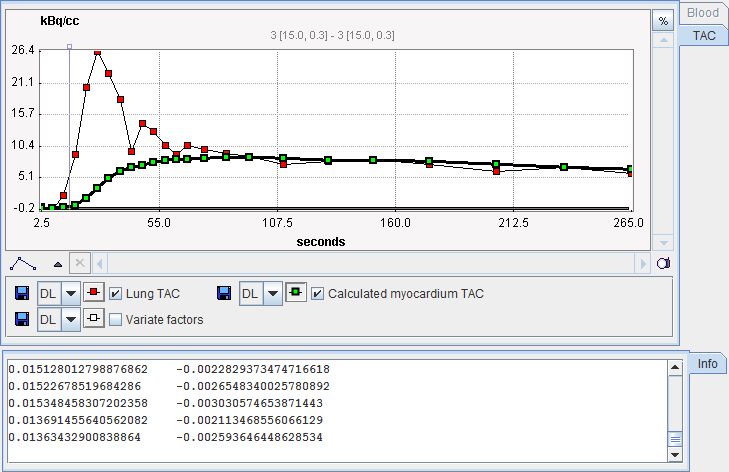
The results of preprocessing is shown in the Results panel.
Model Configuration

Myo |
The myocardium factor images which should represent an anatomical image of myocardium. |
BV |
Blood volume factor images which should show the blood volume. |