![]()
![]()
![]()
![]()
|
|
|
|
|
Wu and Carson [1] aimed at making the SRTM basis function approach even more robust and called it Simplified Reference Tissue Model 2 (SRTM2). They noted that with SRTM k2' is calculated with each pixel TAC, although the same reference TAC is used for all pixels. Therefore they implemented a two-step approach:
The operational equation of the SRTM was re-written to allow for fixing of k2'. This is relevant for parametric mapping because the model in each pixel TAC uses the same reference TAC and therefore should employ the same k2'. Defining the ratio of tracer delivery R1 as K1/K1' and the binding potential BPND as k3/k4, the following operational equation can be derived for the measured TAC in a receptor-rich region:
![]()
The three unknowns R1, k2 and k2a in this equation can be fitted using nonlinear regression techniques. The binding potential can then be calculated as
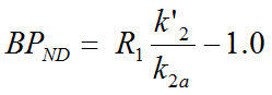
The current PXMOD implementation BPnd (Wu SRTM2 Ref) allows estimating k2' as the median of all k2' values calculated by SRTM in a defined VOI. IN contrast to [1] and unweighted fitting is employed. Note that there is a deprecated version of the model which used the k2' obtained from regional fitting.
Acquisition and Data Requirements
Image Data |
A dynamic PET data set imaging a receptor tracer which behaves kinetically similar to a 1-tissue compartment model. |
TAC 1 |
TAC from a receptor-rich region (such as basal ganglia for D2 receptors). |
TAC 2 |
TAC from a receptor-devoid reference region (such as cerebellum or frontal cortex for D2 receptors). |
VOI |
VOI definition of the brain excluding the reference tissue which can (optionally) be used for getting an estimate of k2'. Recommendation is to use the relevant brain regions outside the reference tissue. |
Model Preprocessing
Two regional TACs (TAC1 and TAC2) as well as a VOI are needed for Model Preprocessing.
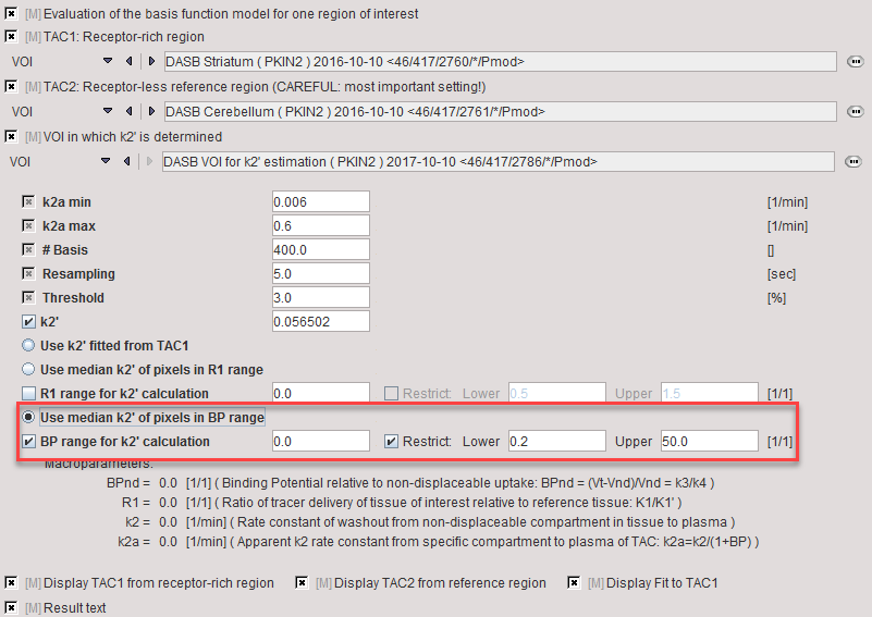
In the lower part the following parameters are configured:
k2a min |
Minimal value of k2a (slowest decay of exponential). |
k2a max |
Maximal value of k2a (fastest decay of exponential). |
# Basis |
Number of basis functions between k2a min and k2a max. Note that increments are taken at logarithmic steps. This number is directly proportional to processing time. |
Resampling |
Specifies the interval of curve resampling which is required for performing the operation of exponential convolution. Resampling should be equal or smaller than the shortest frame duration. |
Threshold |
Discrimination threshold for background masking. Will not have an effect if a mask image has been defined. |
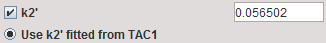
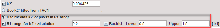
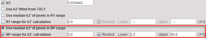
k2' |
k2 of the reference tissue. It can be specified in four different ways:
Note that checking of the ranges is not required for the procedure, only for changing the restriction range. |
BPnd |
Estimated binding potential (= k3/k4 according to the underlying model). |
R1 |
Ratio of tracer delivery in each pixel relative to the reference tissue (R1=K1/K1'). |
k2 |
Estimated rate constant k2. |
k2a |
k2a value which provides the best least squares fit in each voxel. |
The result of the regional fit during Model Preprocessing is shown in the Results panel for inspection.
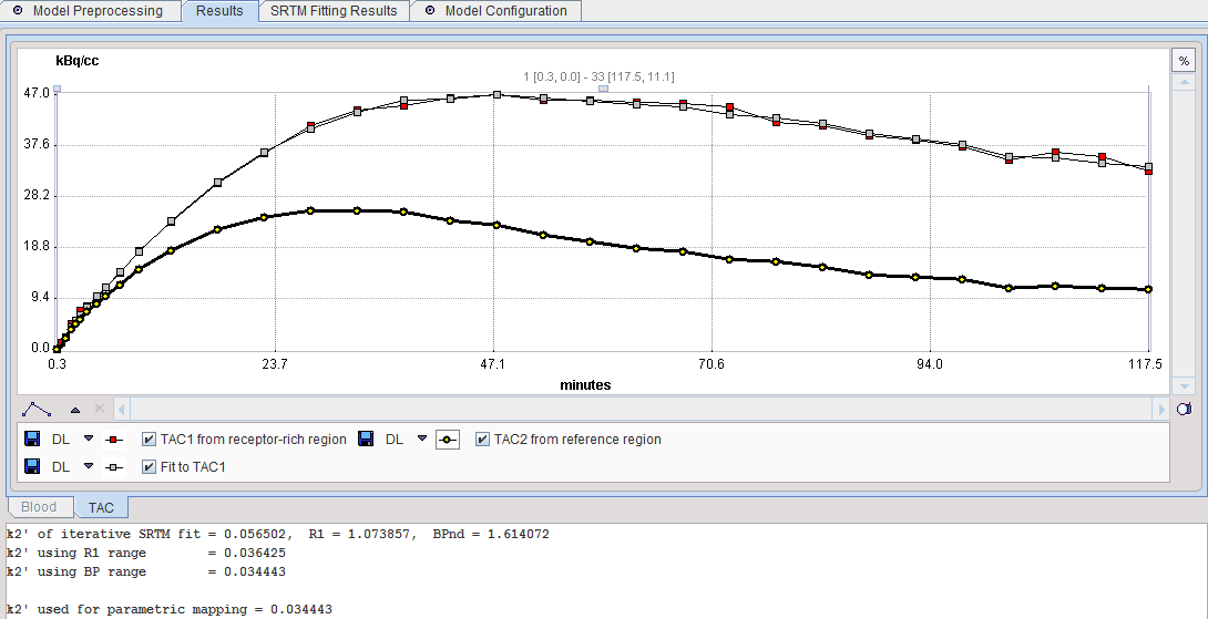
Note the lower text section which lists the k2' results of all three procedures as well as the k2' configured for the pixelwise analysis.
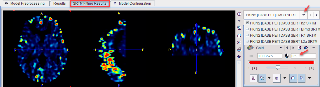
The model features an additional panel SRTM Fitting Results which allows inspecting the SRTM parametric maps with in the VOI. The parameter can be switched in the upper right. Due to outlier results it may be required to manually enter a physiologic value as the upper image threshold as illustrated below.
Model Configuration
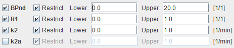
BPnd |
Estimated binding potential (BPnd= k3/k4 according to the underlying model). |
k2 |
Estimated efflux rate constant k2 . |
R1 |
Ratio of tracer delivery in each pixel relative to the reference tissue (R1=K1/K1'). Therefore the map often has a similar appearance to a perfusion image. |
k2a |
k2a value which provides the best least squares fit. |
Note: The k2a parametric map should be checked in the initial setup of a processing protocol. The estimated k2a values should not be truncated by too narrow k2a min and k2a max values.
Reference
1. Wu Y, Carson RE: Noise reduction in the simplified reference tissue model for neuroreceptor functional imaging. J Cereb Blood Flow Metab 2002, 22(12):1440-1452. DOI