PERCIST-like VOIs
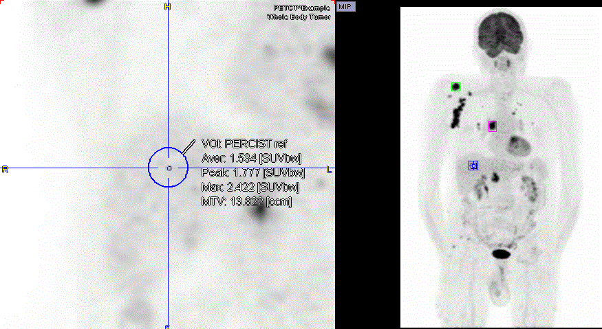
Instead of basing iso-contouring on the values in the lesion itself or an absolute SUV, the threshold can be obtained from the uptake in the reference tissue. The PERCIST (PET Response Criteria in Solid Tumors) [1,2] obtains the background activity from a 3-cm diameter sphere placed in the right side of the liver, midway between the dome and inferior margin, excluding central ducts and vessels. If the liver is diseased, background is to be measured in the descending thoracic aorta (cylinder: 1cm diameter, 2cm long, avoiding wall).
The procedure for a PERCIST-conformant assessment is as follows:


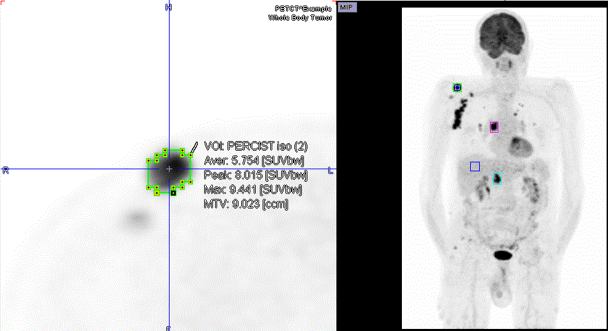
For each measurable lesion a corresponding entry will appear in the VOIs list named PERCIST iso followed by a number through round brackets, e.g. (3), indicating the order of the outlining.
Each lesion can be documented per screen capture. Note the Peak value, which is the relevant outcome for PERCIST.
Sorting of Segmented Lesions
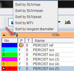
Once the lesions have been segmented, they can be ordered according to different criteria, such as size, MTV, SUVmean, SUVpeak, SUVmax. In this way, the most relevant lesions are easily brought to the top of the list for further evaluation and documentation.

In contrast to RECIST, which employs the largest diameter in the measurement plane to quantify lesion size, PMOD supports the longest oblique diameter in three dimensional space.